At the office of William L. Krell, DDS, MAGD, we invest in advanced diagnostic tools so every treatment begins with a clear, three-dimensional picture of your oral anatomy. Cone-beam computed tomography (CBCT) gives our clinical team detailed, distortion-free images of teeth, jawbone, nerves, and sinuses—information that simply isn’t available from conventional two-dimensional X-rays alone.
Using CBCT allows us to plan care with greater confidence and fewer surprises. The scans are fast and focused on the area of interest, helping us design treatment plans that are safer, more predictable, and better tailored to each patient’s anatomy and goals.
CBCT is a specialized imaging technology that captures a volumetric dataset of the head and neck in a single, short scan. Unlike standard dental X-rays that compress complex anatomy into a flat image, CBCT preserves spatial relationships and depth. This makes it possible to measure bone dimensions, evaluate tooth root form, and visualize structures that might otherwise be hidden or misinterpreted.
The level of anatomical detail from CBCT supports clearer diagnoses of conditions such as impacted teeth, root fractures, and complex root canal systems. Clinicians can rotate, slice, and zoom into the dataset to view cross-sections at precise angles, which improves diagnostic accuracy and reduces the need for exploratory procedures.
Because CBCT data is digital, it integrates smoothly with other treatment technologies—implant planning software, surgical guides, and digital crowns—creating a seamless workflow from diagnosis through restoration. This compatibility shortens treatment timelines and helps align surgical and prosthetic goals from the outset.
Detailed three-dimensional imaging transforms generic treatment into individualized care. For complex cases—such as multi-rooted teeth, challenging extractions, or reconstructive needs—CBCT gives the information necessary to anticipate anatomic variations and plan interventions accordingly. This reduces intraoperative uncertainty and allows for more conservative approaches when appropriate.
In restorative dentistry, CBCT helps assess the volume and quality of jawbone, identify the precise location of nerves and sinus cavities, and determine the best orientation for implants or grafts. For endodontic procedures, it reveals root canal anatomy and periapical pathology in a way that 2D images cannot, informing treatment decisions that preserve natural tooth structure when possible.
The result is a treatment plan that reflects your unique anatomy and treatment priorities—whether the goal is long-term function, improved comfort, or a natural-looking restoration. CBCT data also makes interdisciplinary coordination simpler, enabling clearer communication between specialists when multiple providers are involved.
Successful implant care depends on accurate assessment of bone height, width, and density as well as safe distance from nerves and sinuses. CBCT provides the precise measurements needed to select implant size and position, reducing the risk of complications and improving prosthetic outcomes. When combined with digital planning tools, CBCT scans enable placement that is prosthetically driven—meaning the final restoration guides the surgical approach.
For many patients, CBCT-guided planning leads to minimally invasive surgery. By identifying optimal implant sites and avoiding critical anatomy, we can often reduce surgical time and trauma. In some cases, CBCT datasets are used to fabricate surgical guides that translate a virtual plan into highly accurate clinical execution.
Whether placing a single implant or rebuilding a full arch, CBCT helps us build a predictable pathway from temporary restoration to long-term function, so you can understand the stages of treatment and the rationale behind each clinical decision.
Radiation exposure is an important consideration with any imaging modality. Modern CBCT systems used in dental offices are designed for dental-dose protocols: focused fields of view, short scan times, and optimized settings that capture necessary detail while minimizing exposure. Our team follows established safety guidelines and selects the smallest field of view appropriate for the diagnostic task.
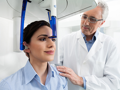
The scanning process itself is quick and noninvasive. Patients typically remain seated or standing while the machine rotates around the head for a matter of seconds. There is no need for intrusive sensors or uncomfortable positioning; most people find the experience straightforward and well tolerated.
As with any diagnostic tool, we recommend imaging only when the expected clinical benefit outweighs any potential risk. When imaging is recommended, we will explain why CBCT is the best option for your case and how the information will be used to improve outcomes.
A CBCT appointment begins with a brief review of your health history and the clinical questions we aim to answer. If a scan is indicated, our trained staff will position you, review safety precautions, and run the quick scan. The raw data is then processed into a 3D dataset that the clinician reviews using specialized software.
During follow-up, we’ll review key findings with you in clear, visual terms—showing cross-sections and measurements and explaining how those details influence treatment choices. Because CBCT images are digital, we can annotate and compare them to treatment plans, models, or other diagnostic records to help you see the whole picture.
Using CBCT, our goal is to reduce uncertainty, shorten treatment timelines when possible, and help you make well-informed decisions about your care. The imaging process is just one step in a comprehensive plan designed to restore health, function, and confidence.
In summary, cone-beam computed tomography is a powerful diagnostic tool that enhances our ability to diagnose, plan, and execute dental treatments with greater precision. At the practice of William L. Krell, DDS, MAGD, we use CBCT judiciously to support safer, more predictable care across a wide range of clinical needs. Contact us to learn more about how CBCT may play a role in your treatment plan and to discuss whether advanced imaging is appropriate for your situation.
CBCT is a three-dimensional imaging technique that captures a volumetric dataset of the head and neck in a single, short scan. Unlike conventional two-dimensional X-rays, CBCT preserves spatial relationships and depth so clinicians can measure bone dimensions and evaluate tooth root form accurately. The scan produces distortion-free cross-sections that can be rotated, sliced, and zoomed to inspect anatomy from multiple angles. This capability reduces diagnostic ambiguity and provides information that 2D images often cannot convey.
The detailed 3D data set from CBCT integrates with digital treatment tools such as implant planning software and CAD/CAM systems, creating a continuous workflow from diagnosis through restoration. Because the images are digital, they can be annotated, compared, and shared securely with specialists when coordinated care is required. The technology therefore supports more precise, predictable treatment planning and fewer intraoperative surprises. Clinicians use CBCT selectively for cases where 3D information will influence clinical decisions.
CBCT is particularly useful for diagnosing impacted teeth, complex root canal anatomy, vertical root fractures, and periapical pathology that are difficult to visualize on 2D films. It also aids in evaluating bone defects, cysts, sinus conditions, and temporomandibular joint anatomy when clinical signs suggest deeper involvement. Orthodontic and airway assessments can benefit from volumetric views when three-dimensional relationships are relevant to treatment. In trauma cases, CBCT helps determine fracture extent and fragment position more reliably than standard radiographs.
On the surgical side, CBCT supports implant placement, alveolar ridge assessment, and bone graft planning by showing bone volume and the location of vital structures such as nerves and sinuses. It is valuable for preoperative planning of complex extractions and reconstructive procedures where precise anatomic detail reduces risk. The images also improve communication between general dentists and specialists, leading to coordinated, patient-centered plans. Ultimately, CBCT is selected when its additional information will change or refine treatment choices.
CBCT provides precise measurements of bone height, width, and density and identifies the spatial relationship of nerves and sinus cavities, information that is essential for selecting implant size and position. These measurements help clinicians avoid critical anatomy and determine whether bone grafting or alternative approaches are necessary. With accurate three-dimensional visualization, treatment can be prosthetically driven so the final restoration, not just available bone, guides implant placement. This approach reduces the likelihood of complications and improves the predictability of prosthetic outcomes.
When combined with implant planning software, CBCT datasets can be used to design and fabricate surgical guides that translate a virtual plan to the clinical setting with high accuracy. Guides can shorten surgical time and support minimally invasive techniques by limiting tissue trauma and the need for broad exposure. The detailed plan also improves communication with dental laboratories and specialists involved in restoration, enabling smoother transitions from implant placement to final prosthesis. Overall, CBCT-led planning enhances safety, function, and long-term success for implant cases.
Modern dental CBCT systems are designed for dental-dose protocols that use focused fields of view, short scan times, and optimized exposure settings to capture the necessary detail while minimizing radiation. Clinicians follow the ALARA principle (as low as reasonably achievable) and select the smallest field of view and lowest acceptable settings that meet the diagnostic need. Compared with a full medical CT scan, dental CBCT typically delivers a substantially lower dose, though it may be higher than a single intraoral radiograph depending on the selected protocol. The decision to image with CBCT balances the diagnostic benefit against any potential exposure risk.
CBCT is not recommended routinely for every visit and should be used only when it will directly inform diagnosis or treatment. Special considerations apply for pregnant patients and children, and clinicians discuss alternative imaging or deferment when appropriate. Your provider will review the reason for the scan and explain how the CBCT findings will be used to improve care before imaging is performed. In all cases, careful justification and protocol selection are primary safeguards.
A CBCT appointment typically begins with a brief review of your medical and dental history and a discussion of the clinical question the scan is intended to answer. During the scan you will be positioned seated or standing while the machine rotates around your head; the actual acquisition usually takes only a few seconds. There are no intraoral sensors or invasive devices required and most patients find the experience quick and well tolerated. Staff will take routine safety precautions and select the appropriate field of view for the area of interest.
After acquisition, the raw data are processed into a 3D dataset that the clinician reviews with specialized software. During a follow-up, your provider will show key cross-sections and measurements and explain how those details influence treatment recommendations. The digital images can be annotated, compared with other records, and shared with any specialists involved in your care. This review helps you understand the reasons for recommended procedures and the expected steps in your treatment plan.
In endodontics, CBCT is invaluable for revealing complex canal morphology, detecting additional or accessory canals, and identifying vertical root fractures that may be missed on 2D films. It also provides clear visualization of periapical pathology and the spatial relationship of roots to surrounding structures. This information helps determine whether a tooth is amenable to nonsurgical root canal therapy or whether surgical intervention may be required. By clarifying anatomic complexities, CBCT supports treatment strategies aimed at preserving natural tooth structure when possible.
CBCT can guide decision-making in retreatment cases by revealing previously undetected anatomy or persistent infection pathways. It also assists in assessing the proximity of critical anatomy before apical surgery and in planning surgical approaches with greater precision. While not required for every endodontic case, CBCT is used selectively when the expected diagnostic benefits will influence the course of treatment. The resulting clarity can reduce the need for exploratory procedures and improve the likelihood of a favorable outcome.
Yes. Because CBCT creates three-dimensional images, it can reveal buccal or lingual bone defects, small periapical lesions, root fractures, and resorptive defects that may be obscured by overlapping structures on conventional radiographs. Cross-sectional views eliminate the compression of complex anatomy into a flat plane, making subtle findings easier to identify. CBCT also provides precise measurements and orientation that help distinguish true pathology from artifacts or normal anatomic variations. This enhanced detection can change diagnosis and influence treatment selection in many cases.
That said, not every clinical question requires 3D imaging, and CBCT should be reserved for situations where additional anatomic detail will change management. Clinical examination and traditional radiographs remain important first-line tools, and CBCT is used when those modalities leave uncertainty. Your clinician will interpret CBCT findings in the context of clinical signs and symptoms to form a comprehensive diagnosis. Integrating all available information yields the most reliable assessment.
CBCT has limitations, including relatively low soft-tissue contrast compared with medical CT or MRI, and susceptibility to artifacts from metal restorations that can obscure adjacent structures. Large-volume scans may increase exposure and are not necessary if a smaller field of view will answer the clinical question. CBCT is also less useful for purely soft-tissue diagnoses where other imaging modalities are better suited. For routine screening or uncomplicated cases, conventional radiographs often provide sufficient information with lower exposure.
CBCT is contraindicated when the imaging outcome will not affect patient management or when alternative imaging adequately addresses the clinical concern. Additional caution is advised for pregnant patients and for choosing pediatric protocols; these choices are made after careful clinical justification. Ultimately the clinician determines whether CBCT is the appropriate diagnostic tool based on the expected benefit, available alternatives, and patient-specific factors. Clear communication ensures the imaging plan aligns with each patient’s needs.
CBCT datasets are fully compatible with implant planning and CAD/CAM software, allowing clinicians to merge volumetric anatomical data with digital impressions and prosthetic designs. This integration enables prosthetically driven implant planning where the desired restoration determines implant position and angulation. The resulting virtual plan can be used to design and mill surgical guides that transfer the plan accurately to the clinical setting, improving placement precision. Digital workflows that include CBCT shorten treatment timelines by coordinating surgical and restorative steps more effectively.
Beyond implants, CBCT data support fabrication of custom grafts, guides for complex extractions, and interdisciplinary planning between general dentists, oral surgeons, and laboratory technicians. The shared digital files allow teams to review the same annotated images and measurements, improving consistency and predictability. Those efficiencies translate into clearer treatment staging and fewer unexpected intraoperative changes. When used judiciously, CBCT is a central element of modern, digitally driven dental care.
At the office of William L. Krell, DDS, MAGD, CBCT is used selectively to clarify complex anatomy and support treatment planning for implants, endodontic cases, surgical procedures, and reconstructive needs. Our team prioritizes the smallest field of view and optimized settings that answer the clinical question while minimizing exposure. The 3D images are incorporated into a digital workflow for planning and, when appropriate, for designing surgical guides that translate virtual plans into accurate clinical execution. This approach helps reduce intraoperative uncertainty and supports predictable restorative outcomes.
Following image acquisition, clinicians review annotated cross-sections and measurements with patients in clear, visual terms so treatment recommendations are well understood. CBCT images are also used to coordinate care with specialists when multiple providers are involved, ensuring treatment goals remain aligned. By combining CBCT data with clinical examination and other diagnostic records, our team aims to develop individualized plans that prioritize safety, function, and long-term stability.

Get in touch with us today!