Digital radiography replaces traditional film with electronic sensors and computer processing to capture images of your teeth, jaw, and supporting structures. Rather than waiting for chemical development, the images appear on a monitor within seconds, giving both patient and clinician immediate insight. This shift from film to digital has reshaped how dentists evaluate oral health—making diagnosis faster, recordkeeping simpler, and communication with specialists more efficient.
At the office of William L. Krell, DDS, MAGD, digital radiography is part of a broader commitment to using contemporary tools that enhance clinical excellence without compromising patient comfort. The sensors used are designed to sit comfortably in the mouth, while the software allows the dental team to adjust contrast, zoom in on areas of concern, and compare images from different appointments. These capabilities help the dentist spot problems earlier and plan treatment more precisely.
Because digital images are captured electronically, they integrate directly into your dental record and can be archived, retrieved, and backed up easily. That means fewer lost films, clearer longitudinal tracking of changes over time, and a smoother workflow for both routine checkups and more complex restorative or implant treatment planning.
One of the most appreciated benefits of digital radiography is its focus on patient safety. Digital sensors require less radiation exposure than many traditional film techniques, and modern equipment is calibrated to capture the necessary diagnostic detail with minimal dose. For patients who require frequent monitoring—such as those undergoing extensive restorative work—this reduction in exposure is an important consideration.
Comfort is another practical advantage. Sensors come in a variety of shapes and sizes to better accommodate different mouths, which typically makes the image-taking process quicker and less intrusive. Because images appear instantly, the team can confirm a clear shot on the first attempt in many cases, reducing the need for repeat exposures and shortening appointment times.
Digital radiography also eliminates film-developing chemicals and paper waste, which is better for the environment and reduces the laboratory processes that once added steps to care. The result is a cleaner, more streamlined experience that prioritizes safety, convenience, and environmental responsibility.
High-resolution digital images give clinicians a level of visual detail that supports more confident decision-making. Software tools allow dentists to enhance contrast, measure distances, and magnify specific areas—capabilities that help reveal subtle signs of decay, bone loss, or structural anomalies that might be harder to detect on old-style film. The clarity of these images supports early intervention, which can prevent minor issues from becoming major problems.
Beyond everyday diagnostics, digital images play a key role in treatment planning for restorative work and implants. Precise imaging helps determine the correct position for crowns, bridges, and implant components, and can be used alongside other digital technologies—such as CBCT scans and digital impressions—to create a coordinated, predictable treatment pathway. This integrated approach leads to better outcomes and fewer surprises during procedures.
Because images are stored electronically, the dentist can quickly pull up past films to show how a tooth or area of the mouth has changed over time. Visual comparisons are a powerful tool for patient education, enabling a clearer conversation about why a treatment is recommended and what progress or risks exist.
Digital radiography simplifies the administrative side of dentistry by linking images directly to the patient’s electronic chart. That makes retrieval instantaneous, streamlines insurance documentation, and reduces the physical storage burden associated with film. Practitioners can also annotate images and attach them to treatment notes, improving continuity of care across visits and among team members.
Collaboration with specialists is easier when images are digital. A high-quality file can be shared securely with an oral surgeon, periodontist, or orthodontist to facilitate consultation and co-planning without the delays of mailing or transporting physical film. This collaborative workflow shortens the time between diagnosis and implementation of treatment, which benefits patients who need coordinated care.
Finally, because digital files can be encrypted and backed up, they offer advantages in both accessibility and data protection. Security protocols ensure that patient information remains private while still being available to authorized members of the clinical team when needed for efficient, well-informed treatment decisions.
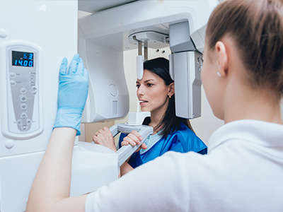
During a routine visit that requires radiographs, a trained member of the dental team will position a small sensor in your mouth and take a series of images that target the areas the dentist needs to evaluate. The process is quick and comfortable for most patients: each exposure lasts only a fraction of a second, and the immediate feedback on-screen allows the clinician to confirm image quality right away. If additional angles are needed, they can be obtained without significant delay.
After the images are captured, the dentist will review them with you on a monitor, pointing out relevant findings and explaining how they relate to your overall oral health. This is an opportunity to ask questions and see the evidence behind any recommended treatment. If needed, digital images can be combined with other technologies—such as intraoral photographs or 3D scans—to provide a comprehensive picture for planning advanced procedures.
Whether you are here for a routine checkup, restorative care, or implant planning, digital radiography helps make the visit more informative and efficient. If you’d like to learn more about how we use imaging to support diagnosis and treatment, please contact us for more information. Our team will be happy to explain how digital tools are part of our commitment to thoughtful, modern dental care.
Digital radiography uses electronic sensors and computer processing to capture images of teeth, jaws, and surrounding structures instead of photographic film. Images appear on a monitor within seconds, eliminating chemical development and allowing immediate review by the clinician and patient. Because the files are digital, they can be adjusted for contrast and magnification to reveal details that might be harder to see on film.
This technology streamlines recordkeeping by integrating images directly into the electronic chart and enables easier archiving and retrieval. Digital workflows also reduce physical storage needs and the risk of lost films, improving continuity of care. In many practices, digital radiography is paired with other digital tools to support coordinated diagnosis and treatment planning.
Digital sensors are designed to capture diagnostic-quality images with lower radiation doses than many traditional film techniques, because they require less exposure to produce a clear image. Modern equipment is calibrated to use the minimum dose necessary and practices follow protocols to limit exposures to what is clinically required. For patients who need frequent monitoring, this reduction in dose is an important safety consideration.
Dental teams also use standard protective measures such as lead aprons and thyroid collars when indicated, and exposures are kept as brief as possible. If you have specific medical concerns or are pregnant, tell your clinician so they can tailor imaging decisions to your needs. The dental team can explain the reasons for any recommended images and discuss alternatives when appropriate.
High-resolution digital images provide clarity that helps clinicians detect subtle signs of decay, bone loss, and other structural changes earlier than may be possible with older film methods. Software tools allow dentists to enhance contrast, measure distances, and magnify areas of interest to support more confident decision-making. Early detection often means less invasive interventions and more predictable outcomes.
Digital files can be combined with three-dimensional scans and digital impressions to create a coordinated treatment plan for restorative work and implants. The ability to compare current and past images side-by-side makes it easier to monitor progression and evaluate treatment response. These capabilities support precision in both routine and complex care.
A trained member of the dental team will position a small electronic sensor in your mouth and take a series of brief exposures targeted to the areas the dentist needs to evaluate. Each exposure lasts only a fraction of a second, and the image appears almost instantly on a monitor so the team can confirm quality right away. Because of the immediacy, repeat exposures are often unnecessary, which shortens the appointment time for most patients.
After images are captured, the dentist will review them with you on-screen and explain any findings and how they relate to your overall oral health. This visual review is an opportunity to ask questions and understand the evidence behind any recommended next steps. If additional views or 3D imaging are needed, the clinician will explain why and how they support the proposed treatment.
Digital radiographs provide precise two-dimensional information about bone levels, root anatomy, and adjacent structures that are essential for planning restorative and implant treatments. When used alongside three-dimensional CBCT scans and digital impressions, these images contribute to accurate measurements and component selection for crowns, bridges, and implants. This multimodal imaging approach helps reduce surprises during surgery and supports predictable prosthetic outcomes.
Digital tools also assist in evaluating the fit and position of restorations and in monitoring healing after implant placement or other surgical procedures. Clinicians can document baseline anatomy and follow postoperative changes, using serial images to assess integration and detect complications early. These capabilities lead to coordinated treatment pathways and clearer communication with the patient and any collaborating specialists.
One of the advantages of digital files is that they can be shared securely and efficiently with specialists, such as oral surgeons, periodontists, or orthodontists, to facilitate consultation and co-planning. High-quality image files can be transmitted electronically in encrypted formats that protect patient privacy and reduce the delays associated with mailing physical film. This rapid exchange supports timely, coordinated care when multiple providers are involved.
Dental practices maintain security protocols and backup procedures to safeguard digital records and ensure authorized access only for members of the clinical team. When you request that images be sent to another provider, the office will follow secure transfer methods and documentation practices. If you have questions about how your records are handled, the dental team can explain their privacy and data protection measures.
Digital radiographs are archived within the patient’s electronic chart, making retrieval instantaneous and enabling side-by-side comparisons of images from different visits. This longitudinal view allows clinicians to track the progression of conditions such as cavities, bone loss, or restorative wear and to evaluate the effectiveness of treatment over time. Consistent imaging protocols improve the value of comparisons and support evidence-based decisions.
Because files can be backed up and duplicated, practices reduce the risk of lost films and maintain reliable records for long-term care. Electronic storage also streamlines administrative tasks such as insurance documentation and referral coordination. If you want to review past images, the dentist can pull them up and walk through any meaningful changes.
Manufacturers produce sensors in a variety of sizes and shapes to better accommodate smaller mouths and to improve patient comfort, which can be especially helpful for pediatric patients. Clinicians use positioning techniques and patient-friendly approaches to minimize discomfort and speed the process, often securing a clear image on the first try. For patients with a sensitive gag reflex, the team can modify positioning or take extraoral images when appropriate.
When intraoral placement is not practical, alternative imaging such as panoramic or cone-beam scans can provide valuable diagnostic information with reduced intraoral manipulation. The dental team will discuss the most appropriate imaging option based on clinical needs and the patient’s comfort. Open communication about past experiences helps the clinician tailor techniques for a more comfortable visit.
Digital radiographs provide detailed two-dimensional views but have inherent limitations in representing three-dimensional anatomy, so they may not reveal the full extent of complex structures or spatial relationships. For surgical planning, implant placement, or assessment of certain pathologies, a three-dimensional CBCT scan can offer more complete information. Image quality can also be affected by positioning, motion, and operator technique, which is why proper training and technique are important.
Some clinical questions require a combination of imaging modalities and clinical examination to reach a definitive diagnosis, and the dentist will recommend additional views when necessary. Digital radiography remains a foundational diagnostic tool, but it is most effective when used as part of a broader diagnostic strategy. Your clinician will explain any recommended imaging so you understand its purpose and value.
The office of William L. Krell, DDS, MAGD combines decades of clinical experience with a commitment to modern technology, using digital radiography as part of a comprehensive diagnostic and treatment workflow. Dr. Krell's status as a Master of the Academy of General Dentistry reflects extensive post-graduate training, and the practice emphasizes continuing education and careful technique when capturing and interpreting images. This combination of experience and technology supports accurate diagnosis and thoughtful treatment planning.
The dental team follows established safety protocols, secure recordkeeping, and coordinated workflows to share information with specialists when needed, ensuring efficient, well-documented care. Patients are invited to review their images with the clinician so they understand findings and options before treatment. If you have questions about how imaging will be used in your care, the team will explain the rationale and next steps in plain terms.

Get in touch with us today!