An intraoral camera is a compact, pen-sized imaging device that captures high-resolution color photographs and video from inside the mouth. Designed to reach areas a mirror cannot, it provides a magnified, lit view of teeth, gums, restorations, and soft tissues. The device feeds live images to a monitor so both clinician and patient can view the oral environment in real time, improving transparency and understanding during an exam.
Most intraoral cameras use LED illumination and magnification lenses to produce crisp, true-to-color images that reveal surface texture, early decay, fractures, and margin gaps that may not be visible to the naked eye. Digital sensors and modern processing mean the images are clear enough for clinical documentation and for use in communication with dental laboratories and specialists. Because the capture is instantaneous, images can be annotated, archived, and compared over time to monitor progression or healing.
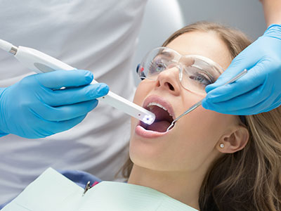
The workflow is straightforward: during a routine exam or a targeted visit, the clinician carefully guides the camera around the mouth while pointing out noteworthy findings on the screen. Patients can ask questions while viewing the same images the dentist sees, which helps demystify diagnoses and creates a shared foundation for discussing treatment options. The process is noninvasive, quick, and often becomes an integral part of a modern dental exam.
By revealing subtle visual cues that may be missed during manual inspection, intraoral cameras enhance the diagnostic process. Early enamel demineralization, tiny cracks in enamel, worn margins on crowns or fillings, and localized soft-tissue changes are easier to identify when magnified and well-lit. This level of detail supports earlier intervention, which can preserve tooth structure and prevent more extensive procedures down the road.
Because the images are stored in the patient’s digital record, clinicians can perform objective comparisons between visits. Serial images provide a reliable method for detecting small changes over time, supporting decisions about observation versus treatment. The ability to freeze-frame and measure areas of concern also aids in clinical judgment and helps prioritize care based on documented progression rather than subjective recall.
In addition to visual detail, intraoral photography facilitates adjunctive assessments. For example, images can be used alongside radiographs and intraoral scanning to corroborate findings and produce a more complete diagnostic picture. This multimodal approach reduces uncertainty and allows clinicians to craft treatment plans that are both conservative and predictable.
Patients often leave a visit with a better understanding of their oral health when they can see what the dentist sees. Visual evidence builds trust and makes explanations about anatomy, disease, and restorative needs more concrete. When patients are shown magnified images of decay, worn restorations, or gum inflammation, they can make informed decisions instead of relying solely on verbal descriptions.
Seeing intraoral photographs also enhances compliance with recommended care. Clear visuals help patients appreciate the rationale behind preventive advice, hygiene recommendations, and the timing of interventions. For anxious patients, the camera can reduce uncertainty by clarifying the extent of a problem and the likely steps for addressing it, which often lowers stress and increases engagement in treatment plans.
Finally, intraoral imaging supports shared decision-making. When clinicians walk patients through documented findings and possible approaches, choices become less abstract. Patients are more likely to follow through with recommended home care and scheduled treatments when they understand the condition and the expected outcomes supported by photographic evidence.
Intraoral cameras are versatile tools used in a wide range of clinical situations. In restorative dentistry they help evaluate margins, detect secondary caries beneath restorations, and document the condition of existing crowns, veneers, and fillings. In periodontal care the camera can capture gingival inflammation, recession, and plaque accumulation—useful for monitoring response to therapy and reinforcing home care techniques.
In implant and prosthetic workflows, photographs from an intraoral camera can be shared with dental laboratories to convey shade, tissue contours, and specific esthetic considerations. The images complement digital impressions and CBCT data, offering surface-level detail that scans sometimes miss. They’re also valuable for surgical follow-up, enabling clinicians to record healing stages and maintain a visual record of soft-tissue changes around implants.
Beyond treatment planning, intraoral images are frequently used for documentation and communication. Clear images strengthen referrals to specialists by providing an accurate visual summary of the patient’s condition. They also serve as reliable records when coordinating care across multiple providers or when explaining treatment steps to patients who may seek second opinions.
Using an intraoral camera is simple and comfortable. During your appointment, the clinician or assistant will position the device inside the mouth for brief moments to capture the necessary views. Each pass typically takes only seconds; if needed, a few additional images are taken to ensure comprehensive coverage. The process is noninvasive, does not produce any sensation beyond mild awareness of the device, and is suitable for patients of all ages.
Captured images are immediately integrated into the patient’s digital chart where they can be annotated, organized, and compared alongside radiographs, digital impressions, and CBCT scans. This integration supports a cohesive, technology-driven workflow that improves care coordination and record-keeping. The visual records are also helpful when discussing esthetic goals, restorative options, and preventive strategies in a way that’s easy to revisit at future visits.
At our state-of-the-art Houston practice, intraoral cameras are one component of a broader commitment to modern diagnostic tools and patient-centered communication. The technology enhances clinical accuracy while giving patients a clear, evidence-based perspective on their oral health. If you have questions about intraoral imaging or how it may be used during your next visit, we’re happy to explain the process and show examples during your exam.
In summary, intraoral cameras are a powerful diagnostic and communication tool that bring clarity and precision to dental care. By producing high-quality images that become part of the permanent record, this technology supports early detection, better patient understanding, and more predictable treatment outcomes. To learn more about how intraoral imaging is used in our practice or to discuss what to expect at your next visit, please contact us for additional information.
An intraoral camera is a compact, pen-sized imaging device that captures high-resolution color photographs and short video clips from inside the mouth. It uses LED illumination and magnifying optics to reveal surface texture, small defects, and soft-tissue detail that can be difficult to see with the naked eye. Images are transmitted instantly to a chairside monitor so clinician and patient can view the oral environment in real time.
The device’s digital sensors and processing produce true-to-color images suitable for documentation, treatment planning, and communication with laboratories or specialists. Captures are instantaneous, which allows clinicians to freeze-frame, annotate, and archive views for comparison over time. Because the workflow is quick and noninvasive, intraoral cameras are easily incorporated into routine exams and targeted visits.
Intraoral cameras enhance visualization of early enamel demineralization, hairline cracks, worn margins on restorations, and localized soft-tissue changes that may be missed during manual inspection. This magnified detail supports earlier detection and more conservative intervention when appropriate. High-quality images also create a clear visual record that helps track progression or healing across successive visits.
Beyond detection, the photographs improve interdisciplinary communication by conveying surface-level detail that complements radiographs and scans. Images help the clinical team and dental laboratory collaborate on esthetic and restorative decisions with greater precision. Overall, intraoral imaging contributes to more predictable, evidence-driven care.
Intraoral photographs reveal subtle visual cues—such as small fractures, margin gaps, and early decay—that can influence diagnostic judgment and the timing of treatment. The ability to freeze-frame, magnify, and measure areas of concern aids objective assessment rather than relying solely on subjective recall. Serial images stored in the chart enable clinicians to compare changes over time and make decisions based on documented progression.
When combined with radiographs, CBCT scans, and intraoral scans, photographic detail completes a multimodal diagnostic picture. This reduces uncertainty and helps prioritize conservative approaches when appropriate. The combined data set supports detailed treatment plans that are both clinically sound and easier to explain to patients.
Using an intraoral camera is generally comfortable and noninvasive; the device is designed for brief, gentle passes inside the mouth and typically causes no sensation beyond mild awareness. Most views are captured in seconds, and additional images are taken only as needed to ensure comprehensive documentation. Because the device is small and maneuverable, it is suitable for adults, adolescents, and most pediatric patients.
Clinicians and assistants adapt technique for patients who have a strong gag reflex, limited opening, or special needs, and staff explain each step to minimize anxiety. The short duration and clear benefit of seeing on-screen images often make the process reassuring for patients. If there are specific concerns, the dental team will adjust the approach to maintain comfort and safety.
Captured intraoral images are imported directly into the patient’s digital chart where they can be annotated, organized, and compared alongside radiographs, CBCT data, and digital impressions. This integration creates a cohesive record that supports longitudinal assessment and treatment planning. Clinicians can easily retrieve prior images to demonstrate change or healing at follow-up visits.
Images exported for laboratory communication or specialist referrals provide a clear visual summary of surface features and tissue contours that scans sometimes miss. Because photographs complement three-dimensional data, they improve the accuracy of shade matching, margin evaluation, and esthetic planning. The result is smoother coordination across providers and more predictable restorative outcomes.
In cosmetic and restorative workflows, intraoral cameras document the condition of enamel, existing restorations, and surrounding soft tissue to guide conservative treatment choices. Photographs help evaluate margins, detect secondary caries, and record color and texture details important for matching restorations. They also serve as a visual reference when discussing options such as veneers, crowns, or teeth whitening.
When communicating with dental laboratories, intraoral images convey shade nuances and gingival contours that improve the fit and esthetic integration of restorations. Postoperative and follow-up photos document healing, restoration integrity, and patient outcomes over time. This visual documentation supports quality control and helps ensure the final result aligns with patient expectations.
An intraoral camera provides detailed surface-level images but does not replace radiographs or CBCT for visualizing internal tooth structure, root anatomy, or bone. Radiographic imaging remains essential for detecting interproximal decay, assessing bone levels, and planning implant placement. Each modality has distinct strengths and is selected based on the diagnostic question at hand.
In practice, intraoral photography complements radiographic and three-dimensional imaging by adding color, texture, and fine surface detail. A combined, multimodal approach reduces diagnostic uncertainty and leads to more comprehensive treatment planning. Clinicians determine the appropriate mix of tools for each clinical situation to ensure safe, evidence-based care.
Seeing clear, magnified images of their own teeth and gums helps patients understand diagnoses and the rationale for recommended care. Visual evidence turns abstract descriptions into concrete observations, which supports informed consent and shared decision-making. When patients view annotated images alongside the clinician’s explanation, they are better equipped to weigh options and ask targeted questions.
Photographs also enhance adherence to preventive advice by illustrating areas of plaque accumulation, gingival inflammation, or early wear that benefit from improved home care. For anxious patients, the transparency of visual documentation often reduces uncertainty and increases confidence in the proposed plan. Overall, intraoral imaging fosters a collaborative relationship between patient and clinician.
Infection control for intraoral cameras follows standard clinical protocols and manufacturer recommendations, including the use of disposable protective sleeves, thorough surface disinfection, and sterilizable components where applicable. Staff receive training on correct handling, barrier placement, and safe removal to prevent cross-contamination. Routine maintenance and inspection ensure optics and electronics remain reliable for clinical use.
Devices are calibrated and cleaned on a regular schedule to preserve image quality and device longevity, and staff document maintenance as part of office protocols. Captured images are stored securely in the patient’s digital record according to professional recordkeeping standards and privacy policies. These measures protect both patient safety and the integrity of diagnostic images.
At our Houston practice, intraoral cameras are used as a routine part of examinations and targeted consultations to document findings, educate patients, and support treatment planning. The clinician or assistant will position the camera to capture key views while explaining noteworthy observations on the chairside monitor. Images are added to the digital chart so they can be referenced during the same visit or at future appointments.
We use intraoral photographs alongside radiographs, digital impressions, and CBCT data when appropriate to develop a comprehensive, personalized treatment strategy. The visual records also streamline communication with laboratories and specialists when collaborative care is needed. Patients are encouraged to review images with the clinician and ask questions so that treatment decisions are informed and transparent.

Get in touch with us today!